

产品分类
- 抗体 (8710)
- Elisa试剂盒 (8205)
- 生化试剂 (11119)
- 培养基 (1017)
- 标准物质 (2297)
- 蛇毒来源提取试剂 (10)
- 进口常用试剂 (15)
- AB Sciex (2633)
- Axygen (1241)
- BD品牌 (24058)
- Biolegend品牌 (10861)
- 贝克曼Beckman品牌 (30775)
- Bio-Rad品牌 (18421)
- Corning (2045)
- ebioscience (11036)
- Fermentas (1043)
- GE品牌 (16087)
- invitrogen品牌 (48423)
- Pierce (2055)
- Qiagen (1974)
- Roche (1957)
- Thermo Fisher (4972)
- ZYMO RESEARCH (380)
- Spherotech (944)
- 其他分类 (2302)
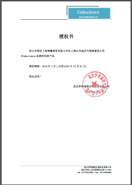
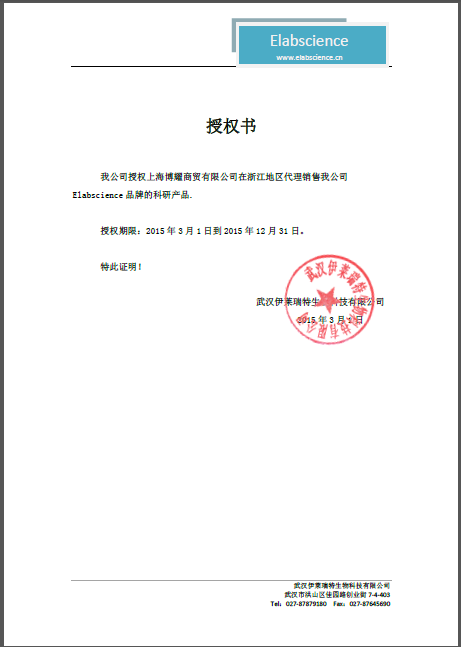
公司荣誉
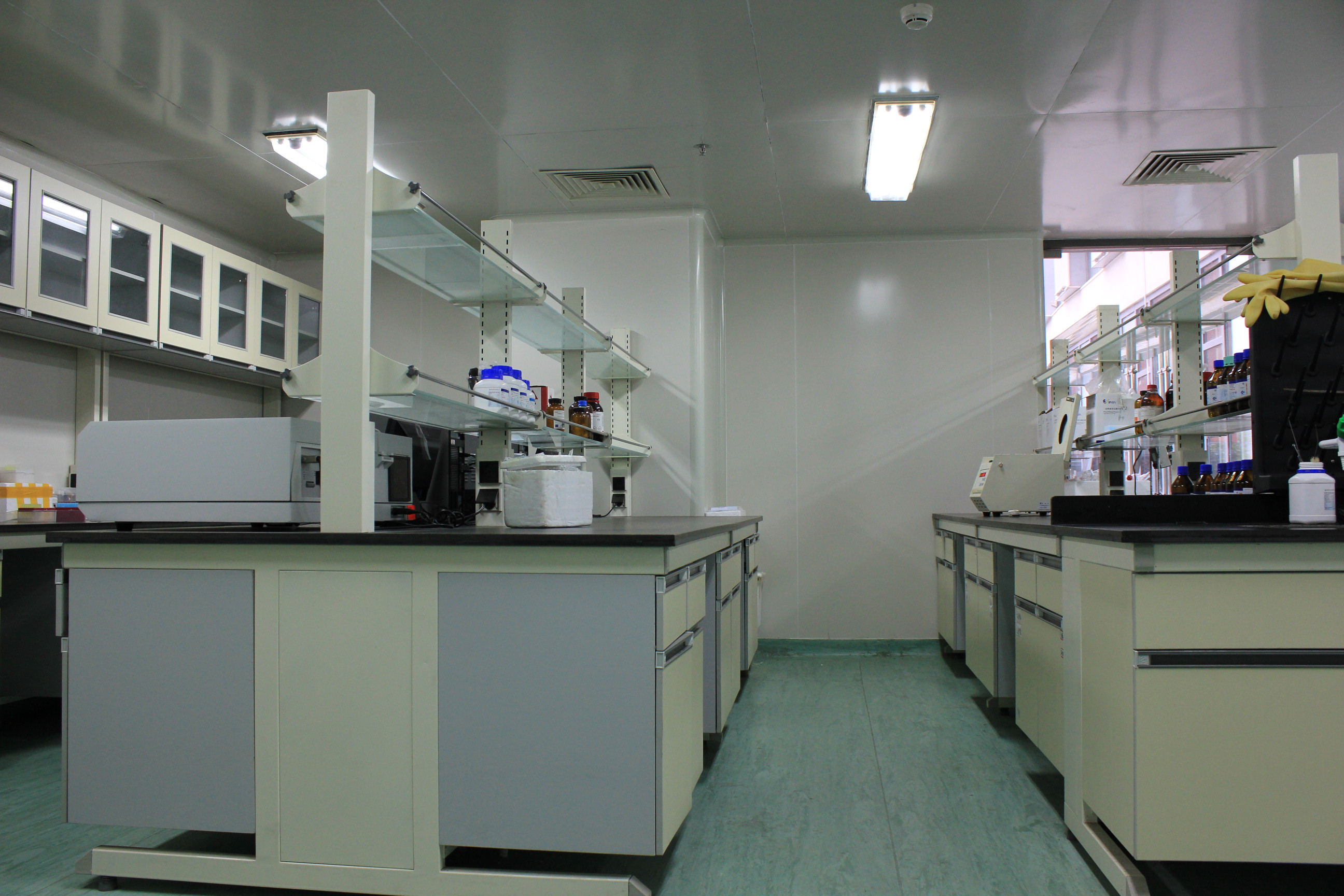
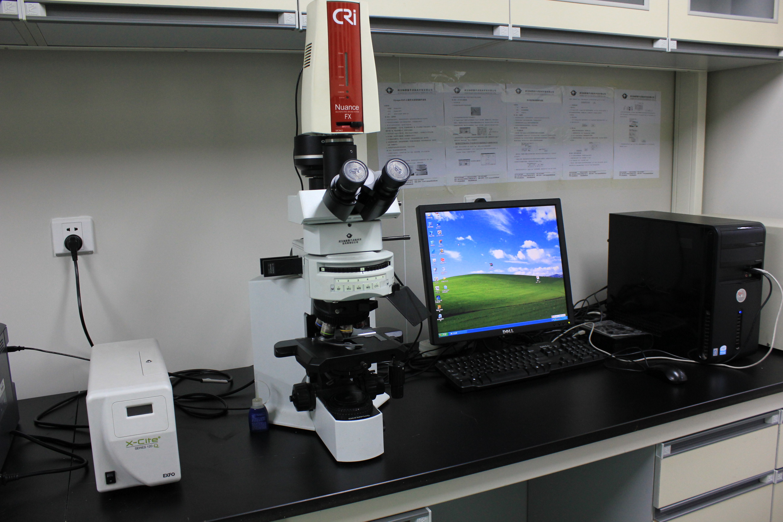
公司图片


公司新闻
![Immunocytochemistry/ Immunofluorescence - Anti-Bax antibody [6A7] (ab5714)](https://img1.dxycdn.com/p/s14/2025/1030/321/8809000143966583891.jpg)
![Immunocytochemistry/ Immunofluorescence - Anti-Bax antibody [6A7] (ab5714)](https://img1.dxycdn.com/p/s14/2025/1030/164/0409931245966583891.jpg)
![Western blot - Anti-Bax antibody [6A7] (ab5714)](https://img1.dxycdn.com/p/s14/2025/1030/886/6792036097966583891.jpg)
](https://img1.dxycdn.com/p/s14/2025/1030/755/6798550901966583891.jpg)